22++ Aortic arch development animation ideas in 2021
Home » Wallpapers » 22++ Aortic arch development animation ideas in 2021Your Aortic arch development animation images are ready. Aortic arch development animation are a topic that is being searched for and liked by netizens today. You can Find and Download the Aortic arch development animation files here. Download all royalty-free photos and vectors.
If you’re looking for aortic arch development animation pictures information connected with to the aortic arch development animation topic, you have pay a visit to the right blog. Our site always provides you with suggestions for seeking the maximum quality video and picture content, please kindly search and locate more enlightening video content and images that match your interests.
Aortic Arch Development Animation. 6 aortic arches have always been spoken about. Animation that illustrates the development of the pericardium from two pleuropericardial folds. An aortic arch is a branch from the arterial aortic sac to the dorsal aorta. These embryonic structures form during the development of the arterial system in intrauterine life.
 Anatomy Branches Of The Aortic Arch Youtube From youtube.com
They are ventral to the dorsal aorta and arise from the aortic sac. The arch gives off three branch vessels the. Better version with labels. Result from errors in the embryologic development of the branchial arches including errors of involution or migration or abnormal persistence of vascular structures. Animation that illustrates the development of the pericardium from two pleuropericardial folds. These embryonic structures form during the development of the arterial system in intrauterine life.
18 Cardiovascular morphogenesis is controlled by mechanisms that are common to all developmental processes.
Better version with labels. Common carotid and initial segments of internal carotid artery. Aortic Arch Vessels Animation and description of the development of the aortic arch vessels aortic sac and descending aorta. The aortic arches or pharyngeal arch arteries previously referred to as branchial arches in human embryos are a series of six paired embryological vascular structures which give rise to the great arteries of the neck and head. They are ventral to the dorsal aorta and arise from the aortic sac. Right brachial aortic arch angiography illustrating a right aortic arch right-sided descending aorta left common carotid artery arising as first branch of the aorta followed by theright common carotid artery and subclavian artery.
 Source: pinterest.com
I based the animat. Right brachial aortic arch angiography illustrating a right aortic arch right-sided descending aorta left common carotid artery arising as first branch of the aorta followed by theright common carotid artery and subclavian artery. Aortic arches are the arteries of pharyngeal arches that arise from aortic sac and terminate into dorsal aorta on each side. This is a revision of the video I made for an Embryology class BYU-I using Apples Motion 4. 6 aortic arches have always been spoken about.
 Source: embryology.ch
The arch gives off three branch vessels the. An aortic arch is a branch from the arterial aortic sac to the dorsal aorta. Maxillary artery in the face 2nd aortic arch. The ascending proximal arch and brachiocephalic artery are derivatives of the aortic sac and truncus arteriosus be aware that some intertextual variation exists. Cell growth cell migration cell death differentiation and adhesion.
 Source: youtube.com
Common carotid and initial segments of internal carotid artery. I made this video for an Embryology class BYU-I using Motion 4. Better version with labels. The ascending proximal arch and brachiocephalic artery are derivatives of the aortic sac and truncus arteriosus be aware that some intertextual variation exists. Aortic and pulmonic valves form The cardiovascular system including the heart blood vessels and blood cells originates from the mesodermal germ layer.
 Source: youtube.com
Aortic and pulmonic valves form The cardiovascular system including the heart blood vessels and blood cells originates from the mesodermal germ layer. The following diagram shows the fate of aortic arches. Maxillary artery in the face 2nd aortic arch. Better version with labels. The arches connect paired dorsal and ventral aortae during development.
 Source: youtube.com
Animation that illustrates the development of the pericardium from two pleuropericardial folds. Aortic arch finalppt 1. The 5th aortic arch forms only a small capillary network and the 6th appears as a prominent capillary network with the early development of the trachea and lungs. Fates of arches 4 and 6. Initially there are five pairs of arches but these undergo structural changes.
 Source: thoracickey.com
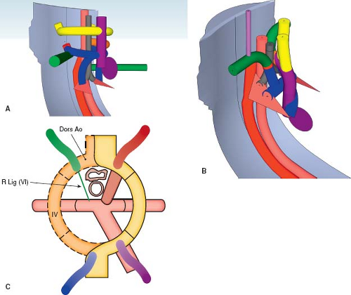
Aortic Arch Vessels Animation and description of the development of the aortic arch vessels aortic sac and descending aorta. The aortic arch is the connection between the ascending and descending aorta and its central part is formed by the left 4th aortic arch during early development. Common carotid and initial segments of internal carotid artery. B Schematic representation of the Edwards hypothetical double arch. It travels in the centre of each pharyngeal arch embedded in mesenchyme.
 Source: slidetodoc.com
The ductus arteriosus connects to the lower part of the arch in foetal life. Cell growth cell migration cell death differentiation and adhesion. B Schematic representation of the Edwards hypothetical double arch. This is a revision of the video I made for an Embryology class BYU-I using Apples Motion 4. I made this video for an Embryology class BYU-I using Motion 4.
 Source: researchgate.net
The arch gives off three branch vessels the. I based the animat. Fates of arches 4 and 6. Aortic arches are the arteries of pharyngeal arches that arise from aortic sac and terminate into dorsal aorta on each side. Animation that illustrates the development of the pericardium from two pleuropericardial folds.
 Source: researchgate.net
It travels in the centre of each pharyngeal arch embedded in mesenchyme. Aortic Arch Anomalies. The 5th aortic arch forms only a small capillary network and the 6th appears as a prominent capillary network with the early development of the trachea and lungs. Initially there are five pairs of arches but these undergo structural changes. Individual vessel development is illustrated.
 Source: pinterest.com
Stapedial artery in middle ear 3rd aortic arch. Normally the aorta ascends in the superior mediastinum to the level of the sternal notch before arching posteriorly and descending in the left hemithorax. These arches have different fates on the left and right sides. Individual vessel development is illustrated. I based the animat.
 Source: youtube.com
Aortic Arch Anomalies. Initially there are five pairs of arches but these undergo structural changes. The aortic arches are formed sequentially within the pharyngeal arches and initially appear. Animation of the developing aortic arches. The arches connect paired dorsal and ventral aortae during development.
 Source: youtube.com
The aortic arch is the connection between the ascending and descending aorta and its central part is formed by the left 4th aortic arch during early development. A cervical aortic arch is a rare anomaly characterized by a high-riding elongated aortic arch Abnormal persistence of 2nd or 3rd primitive aortic arch Ascends into the neck usually on the right. Hospital because of tracheal tubeobstruction. Strong associations of arch anomalies with. The aortic arches are formed sequentially within the pharyngeal arches and initially appear.
 Source: medmovie.com
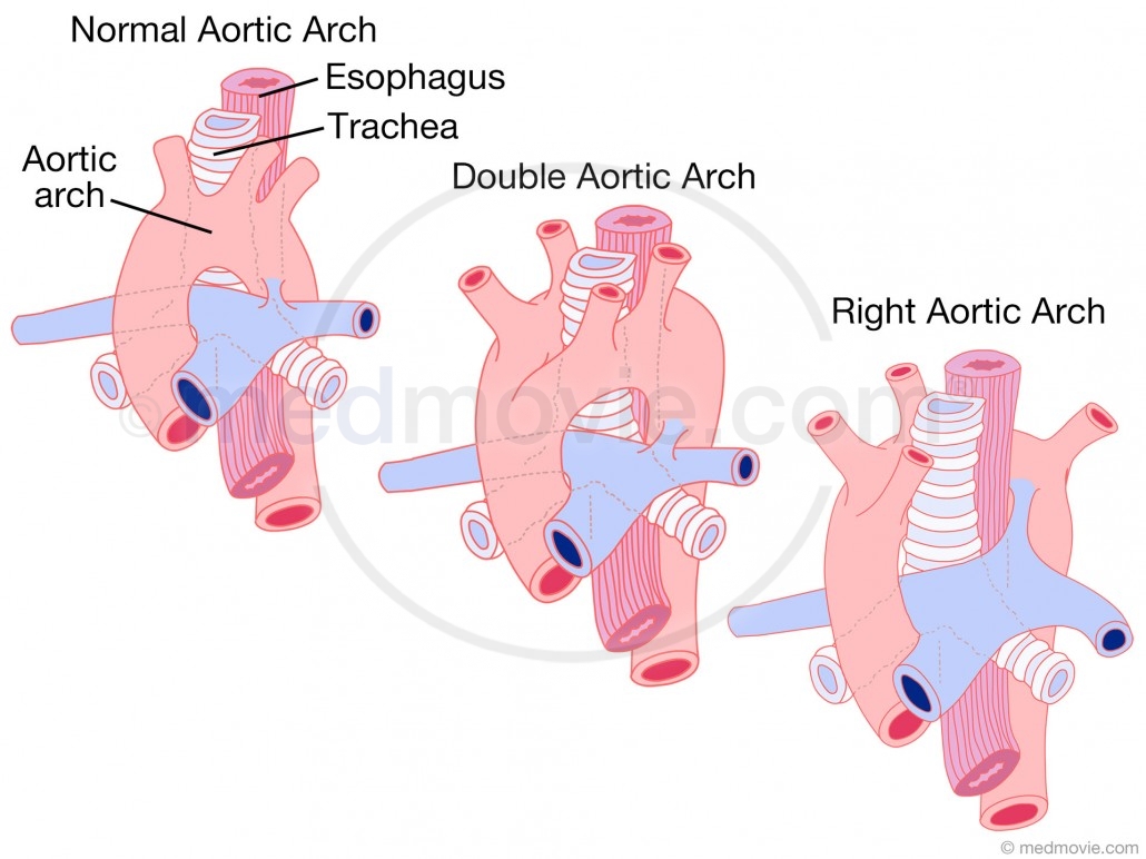
Aortic Arch Vessels Animation and description of the development of the aortic arch vessels aortic sac and descending aorta. The arch gives off three branch vessels the. Normally the aorta ascends in the superior mediastinum to the level of the sternal notch before arching posteriorly and descending in the left hemithorax. Result from errors in the embryologic development of the branchial arches including errors of involution or migration or abnormal persistence of vascular structures. The following diagram shows the fate of aortic arches.
 Source: br.pinterest.com
An aortic arch is a branch from the arterial aortic sac to the dorsal aorta. Individual vessel development is illustrated. A cervical aortic arch is a rare anomaly characterized by a high-riding elongated aortic arch Abnormal persistence of 2nd or 3rd primitive aortic arch Ascends into the neck usually on the right. What is the fate of aortic arches. The aortic arches or pharyngeal arch arteries previously referred to as branchial arches in human embryos are a series of six paired embryological vascular structures which give rise to the great arteries of the neck and head.
 Source: drawittoknowit.com
A Diagram representing six paired branchial arches numbers 16 and an intersegmental artery IA. Also the proximal segment of the 6th arch closest to the aortic sac has a. Initially there are five pairs of arches but these undergo structural changes. Variant anatomy of the aortic arch occurs when there is failure of normal aortic developmentIt results in a number of heterogenous anomalies of the aorta and its branch vessels. What is the fate of aortic arches.
 Source: pinterest.com
These embryonic structures form during the development of the arterial system in intrauterine life. The proximal portion of the right subclavian artery arises from the right fourth aortic arch. Fates of arches 4 and 6. DISCUSSION Anomalies in the development of the aortic arch. 18 Cardiovascular morphogenesis is controlled by mechanisms that are common to all developmental processes.
 Source: aboutkidshealth.ca
Aortic Arch Anomalies. Fates of arches 4 and 6. Right brachial aortic arch angiography illustrating a right aortic arch right-sided descending aorta left common carotid artery arising as first branch of the aorta followed by theright common carotid artery and subclavian artery. Aortic and pulmonic valves form The cardiovascular system including the heart blood vessels and blood cells originates from the mesodermal germ layer. The proximal portion of the right subclavian artery arises from the right fourth aortic arch.
 Source: youtube.com
Initially there are five pairs of arches but these undergo structural changes. The aortic arches are formed sequentially within the pharyngeal arches and initially appear. Right brachial aortic arch angiography illustrating a right aortic arch right-sided descending aorta left common carotid artery arising as first branch of the aorta followed by theright common carotid artery and subclavian artery. The ascending proximal arch and brachiocephalic artery are derivatives of the aortic sac and truncus arteriosus be aware that some intertextual variation exists. Aortic Arch Vessels Animation and description of the development of the aortic arch vessels aortic sac and descending aorta.
This site is an open community for users to do sharing their favorite wallpapers on the internet, all images or pictures in this website are for personal wallpaper use only, it is stricly prohibited to use this wallpaper for commercial purposes, if you are the author and find this image is shared without your permission, please kindly raise a DMCA report to Us.
If you find this site serviceableness, please support us by sharing this posts to your own social media accounts like Facebook, Instagram and so on or you can also save this blog page with the title aortic arch development animation by using Ctrl + D for devices a laptop with a Windows operating system or Command + D for laptops with an Apple operating system. If you use a smartphone, you can also use the drawer menu of the browser you are using. Whether it’s a Windows, Mac, iOS or Android operating system, you will still be able to bookmark this website.